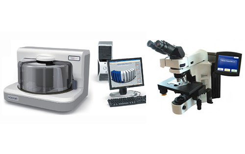
ThinPrepイメージングシステム Duo

細胞診のより確かな判定と効率化を検査室に提供するDual Review™
ThinPrepイメージングシステムDuoは、コンピュータによるイメージング技術と専門技術を有する鏡検者のスキルを融合する“Dual Review(デュアルレビュー)”により、細胞診の確かな判定と鏡検効率の向上を検査室に提供します。
※ThinPrepイメージングシステムDuoは、「ThinPrepイメージングステーション」と「Review Scopeマニュアルプラス」から構成されています。
医療機器届出番号:13B1X10179001008
この製品について
ThinPrepイメージングシステム DuoによるDual Reviewでは、すべてのThinPrep婦人科用プレザーブサイト液(ThinPrep婦人科用バイアル)の検体から作製されたスライドに対して2種類のレビュー、すなわちThinPrepイメージングシステム Duoによるフルレビューと、熟練した細胞検査士によるレビューを行います。あらゆる検体に対してThinPrepイメージングシステム Duoは、熟練した細胞検査士の鏡検をサポートし、精度の高い子宮頸がん検査を可能にします。
ホロジックのThinPrep婦人科用バイアルの検体から作製された標本で使用できるDual Reviewによるスクリーニングは、偽陰性率が低下することが実証されているため2、検査結果の信頼性が高まります。Dual Reviewによるスクリーニングでは、コンピュータ支援による自動化によってスライドをプレスクリーニングし、各細胞および細胞集団のDNA含量に基づいて対象となる細胞を特定します。熟練した細胞検査士が自動顕微鏡で画像化したスライドをレビューする際、鏡検に備えて特に見る必要がある領域がハイライト表示されます。そのため、検査士は、最も重要な点である病変解釈に集中することができます。
性能データ
ThinPrepイメージングシステム Duoは、ThinPrep婦人科用バイアルの検体から作製されたスライドをマニュアルでレビューした場合よりも高い感度と特異度が得られることが示されています。
イメージングシステムの臨床試験結果から、ASC-US以上の検出感度に6.4%という統計学的に有意な増加[95% CI:2.6-10.0]、HSIL以上の検出の特異度に0.2%という統計学的に有意な増加[95% CI:0.06-0.4]が示されました1。一方、Millerらによる2007年の研究からは、ThinPrep婦人科細胞診のスライドをマニュアルでレビューした場合に比べて、HSIL検出率は42%、LSIL検出率は37%増加することが示されました2。
その他、偽陰性、不適正率、ASC-US率の減少も報告されています2。
ホロジックのテクノロジー
ホロジックのDNA染色は、子宮頸部細胞の核染色に用いられています。異常な細胞ではDNAの量が増加し、大きく、または不定形になる傾向があります。また、異常な細胞の核は正常な細胞の核より多くの染色剤を取り込みます。つまり、形状が不規則な細胞や大型の細胞、濃く染色された細胞がある場合、その検体が異常である可能性があります。
ThinPrepイメージングシステム Duoは各標本をスキャンし、対象となる細胞を含む22の視野を特定します。その後、細胞検査士がReview Scope マニュアルプラスを用いてこの22のフィールドをレビューすることができます。細胞検査士がいずれかの視野の細胞について疑わしいと判断した場合は、スライド全体をマニュアルでレビューし、異常な細胞に印を付け、細胞診専門医がさらなるレビューを行うことができます。
参照文献
1.ThinPrep Imaging System Package Insert
2.Miller, et al. Implementation of the ThinPrep Imaging System in a High Volume Metropolitan Laboratory. Diag Cytopath. 2007;35:213-7
参考文献一覧
- Biscotti C, Dawson A, Dziura B, Galup L, Darragh T, Rahemtulla A, Wills-Frank L. Assisted primary screening using the automated ThinPrep Imaging System. Am J Clin Pathol. 2005;123(2):281-7.
- Bolger N, Heffron C, Regan I, Sweeney M, Kinsella S, McKeown M, Creighton G, Russell J, O’Leary J. Implementation and evaluation of a new automated interactive image analysis system. Acta Cytol. 2006;50(5):483-91.
- Chivukula M, Saad RS, Elishaev E, White S, Mauser N, Dabbs DJ. Introduction of the ThinPrep Imaging System (TIS): experience in a high volume academic practice. CytoJournal. 2007;4:6.
- Cibas E, Hong X, Crum C, Feldman. Age-specific detection of high risk HPV DNA in cytologically normal computer-imaged ThinPrep Pap samples. Gynecol Oncol. 2007;104(3):702-6.
- Dawson A. Clinical experience with the ThinPrep Imager System. Acta Cytol. 2006;50(5):481-2. Editorial.
- Dawson AE. The changing face of cervical screening: challenges for the future. Diagn Cytopathol. 2005;33(2):63-4. Editorial.
- Dawson A. Can we change the way we screen?: The ThinPrep Imaging System. Cancer (Cancer Cytopathol). 2004;102(6):340-4.
- Davey E, d’Assuncao J, Irwig L, Macaskill P, Chan SF, Richards A, Farnsworth A. Accuracy of reading liquid based cytology slides using the ThinPrep Imager compared with conventional cytology: prospective study. British Medical Journal. 2007;335(7609):28.
- Davey E, Irwig L, Macaskill P, Chan S, D’Annuncao J, Richards A, Farnsworth A. Cervical cytology reading times: a comparison between ThinPrep Imager and conventional methods. Diagn Cytol. 2007;35(9):550-4.
- Dziura B, Quinn S, Richard K. Performance of an imaging system vs. manual screening in the detection of squamous intraepithelial lesions of the uterine cervix. Acta Ctyol. 2006;50(3):309-11.
- Friedlander MA, Rudomina D, Lin O. Effectiveness of the ThinPrep Imaging System in the detection of adenocarcinoma of the gynecologic system. Cancer (Cancer Cytopathol). 2008;114(1):7-12.
- Holund B, Grinsted P. Screening for cervical cancer in the country of Funen. Status of 25 years of development and experience. Ugeskr Laeger. 2006;168(22):2163-6. Article in Danish.
- Lozano R. Comparison of computer-assisted and manual screening of cervical cytology. Gynecologic Oncology. 2007;104(3):702-6.
- Miller FS, Nagel LE, Kenny-Moynihan MB. Implementation of the ThinPrep Imaging System in a high-volume metropolitian laboratory. Diagn Cytopathol. 2007;35(4):213-7.
- Papillo JL, St. John TL, Leiman G. Effectiveness of the ThinPrep Imaging System: clinical experience in a low risk screening population. Diagn Cytopathol. 2008;38(3):155-60.
- Roberts J, Thurloe J, Bowditch R, Hyne S, Greenberg M, Clarke J, Biro C. A three-armed trial of the ThinPrep Imaging System. Diagn Cytopathol. 2007;35(2):96-102.
- Schledermann D, Hyldebrandt T, Ejersbo D, Hoelund B. Automated screening versus manual screening: a comparison of the ThinPrep Imaging System and manual screening in a time study. Diagn Cytopathol. 2007;35(6):348-52.
- Zhang FF, Banks HW, Langford SM, Davey DD. Accuracy of ThinPrep Imaging System in detecting low-grade squamous intraepithelial lesions. Arch Pahtol Lab Med. 2007;131:773-6.
- Zhao C, Elishaev E, Yuan K, Yu J, Austin RM. Very low human papillomavirus DNA prevalence in mature women with negative computer-imaged liquid-based Pap tests. Cancer (Cancer Cytopathol). 2007;111(5):292-7.


